By Christine Willinger
Measuring blood flow in the brain with MRI may help predict decreases in brain volume due to Alzheimer’s disease (AD), according to a recent study published by Penn Memory Center researchers in the journal Hippocampus.
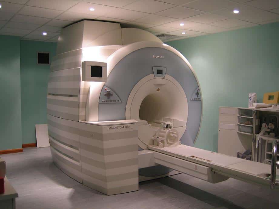
Brain MRI, or magnetic resonance imaging, is a non-invasive imaging method that uses magnetic fields and radio waves to take pictures of the brain. A special type of MRI, called arterial spin-labeled perfusion (ASL) MRI, is able to measure blood flow in different regions of the brain during the imaging process.
Currently, positron emission tomography (PET) imaging is the gold standard for diagnosing AD. However, these scans are expensive and not always covered by insurance, and they involve a small amount of radiation exposure. Could ASL MRI, which is easily incorporated into standard MRI procedures, provide a more affordable alternative for AD diagnosis?
Researchers at PMC studied this very question in a group of participants with and without mild cognitive impairment. They measured cerebral blood flow by ASL MRI while the participants completed a memory task, and then followed up with imaging and clinical testing after a year.
The result: cerebral blood flow during the memory task predicted the rate of decline in brain volume in the hippocampus, which is a key feature of AD progression. The researchers believe that this type of functional imaging using MRI could be more sensitive for detecting early AD than static measurements of structural change.
As they concluded in their paper, “ASL MRI could have important utility in identifying candidates for AD treatment clinical trials likely to display significant progression.” Co-authors on this study included PMC Co-Director David Wolk, MD; neurology researcher Sandhitsu Das, PhD; and study coordinators Arun Pilania, Molly Daffner, and Grace Stockbower.
