By Meghan McCarthy
Author’s Note: This article is part of ongoing coverage of the 2024 Alzheimer’s Association International Conference (AAIC). To view all highlights, please click here.
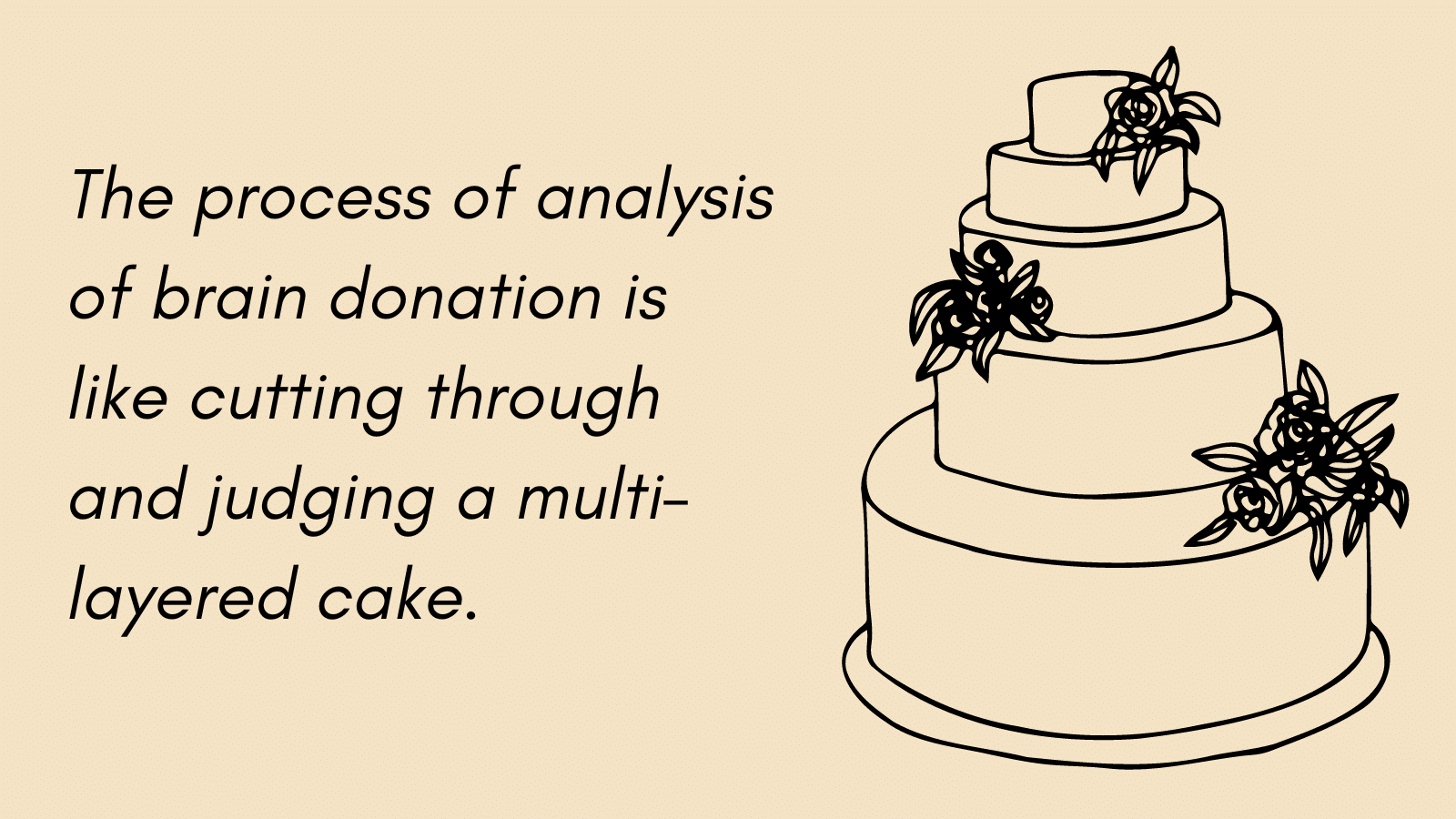
Imagine a tiered, ornately decorated cake. Each tier has a unique flavor, filling, and frosting. The cakes are stacked at various heights and widths and adorned with décor. If you were to slice through each layer, you could distinctly identify each layer’s cake, filling, and frosting. While it would be possible to cut through all tiers together, you could appreciate and evaluate the flavors better by creating slices for the tiers singularly.
The process of analysis of brain donation is like cutting through and judging a multi-layered cake.
Brain Donation
Brain donation and the study of subsequent autopsies are essential components to furthering research in Alzheimer’s disease and related dementias (ADRDs). Once an autopsy is complete, brain tissue is available for further evaluation.
Common methods include magnetic resonance imaging (MRI) and histological evaluation. MRIs allow researchers to have detailed three-dimensional (3D) imaging of brain anatomy. Histology is the study of tissues, and histological examination uses dyes to stain tissue and evaluate cells and their components under a microscope. To study histology after an autopsy, small tissue sections of the brain are stained.
While an MRI would show distinct cake layers and tier shapes, histology would detail the specific flavors and fillings within the cakes.
Penn’s Brain Bank, which is governed by the Center for Neurodegenerative Disease Research (CNDR) and the University of Pennsylvania Alzheimer’s Disease Core Center (ADCC), is one of the largest tissue banks in the country.
Chinmayee Athalye, MS, is a data scientist and bioengineering doctoral student at Penn. She is also a member of the Penn Imagine Computing and Science Laboratory (PICSL).
For scientists such as Athalye, the Brain Bank is a resource to study human brain samples obtained from patients with Alzheimer’s disease, Parkinson’s disease, and other related neurodegenerative dementias and movement disorders.
Over the last few years, researchers at Penn have developed streamlined protocols that allow for efficient processing of the donated brains. They are one of the first teams to look at the whole hemisphere brain histology on this scale.
Athalye’s Registration Pipeline
In her work, Athalye has designed a custom protocol for processing brain autopsies. Her rigorous and analytic process was featured at this year’s AAIC conference and is known as a registration pipeline.
Consider this pipeline similar to a description of a slice of cake If you only had the slice of cake available to you, a registration pipeline would tell you which tier and where this slice was cut from in the original cake.
While Penn has an established protocol for processing an autopsy and storing brain donations, Athalye is interested in what occurs between these steps.
Specifically, she is interested in creating a brain archive that could be used by multiple teams in the future.
The brain has two hemispheres. After an autopsy is performed, traditional Penn protocol assigns one hemisphere for neuropathological analysis. This side will undergo histological staining and have the thickness of the cortex measured. Each histological slide is only 20 microns thick, which is approximately half the thickness of human hair. The team has the ability to scan this at even higher resolutions that allow for assessment of brain tissue on a cellular level.
The second side is evaluated via an MRI.
“It is decent, but measurements are still coming from two different sides of the brain,” Athalye explained.
Compare this to slicing cake from two different tiers: as each have different flavors and components, comparing their slices would be difficult.
To advance these analyses, Athalye and her PICSL team created a pipeline that reviews histology, pathology, and an MRI all from the same side of the brain.
“The lab is a perfect mixture of clinicians, subject matter experts, and completely technical people like myself who work on algorithms,” Athalye said. “I really love all of the collaborative and diverse projects that we work on.”
Specifically, a team of neurologists identified 19 regions of interest (ROIs) within the brain most related to ADRDs. Once marked on a 3D MRI, her team creates a custom 3D mold printed for each brain. This mold includes notches in the mold that are precisely spaced one centimeter apart. Once complete, team generates hundreds of tissue slides for microscopic evaluation.
Envision their 3D mold like cake pan with notches throughout the bottom, guiding an individual on how to best cut the cake.
These tissue sections are then stained based on their ROI and intended analysis. After staining, Athalye applies a machine learning algorithm known as the WildCat. This algorithm is trained to detect biomarkers such as neuronal tangles and the presence of tau proteins, which are commonly found in ADRDs. The WildCat algorithm evaluates each brain tissue section and generates a cumulative biomarker score for each ROI.
Think of the Wildcat like a qualified pastry judge in a bake competition, scoring the cake based on appearance in addition to internal flavors.
“We get a quantitative score for the pathology and through the MRI, have a score for the thickness of the cortex,” Athalye said. “Because our pipeline evaluates ROIs from the same hemisphere, we can draw correlations and establish relationships that are much finer grained.”
Future Directions
This registration pipeline can be customized for many ADRD projects. In her specific work, Athalye is comparing pathological scores of tau with measured cortical thickness.
Beyond her current project, Athalye looks forward to refining the registration pipeline and automating histological analyses as her teams scales up. She is also excited to specifically apply the pipeline to studying white matter hyperintensities, which are MRI abnormalities that are understudied in ADRD research.
“What is great is that our histological slices are meticulously stored and can be continuously analyzed,” Athalye said. “If you want to go into the archive and study them for a different project in the future, it will be possible.”
Athalye has a background in electrical engineering, computer science, and image processing. Specifically, she is interested in applying machine learning technologies to the biomedical field.
To view Athalye’s AAIC poster presentation, please click here.


